A patient arrives with sudden weakness on one side of the body. Another has crushing chest pain after a collision. A third has pain deep in the spine, but the X-ray is silent. In each case, the question is not simply, “Which scan is better?” MRI versus CT scan uses depend on what clinicians suspect is happening, how fast an answer is needed, and what kind of tissue may be hiding the evidence.
The machines can seem equally imposing: a narrow table, a whirring gantry, instructions to hold still while unseen forces build a picture from inside the body. But CT and MRI do not see the same way. One is often built for speed and acute danger. The other is built for contrast, detail, and the slow revelation of soft tissue abnormalities that can remain invisible on other images.
MRI Versus CT Scan Uses: Two Different Investigations
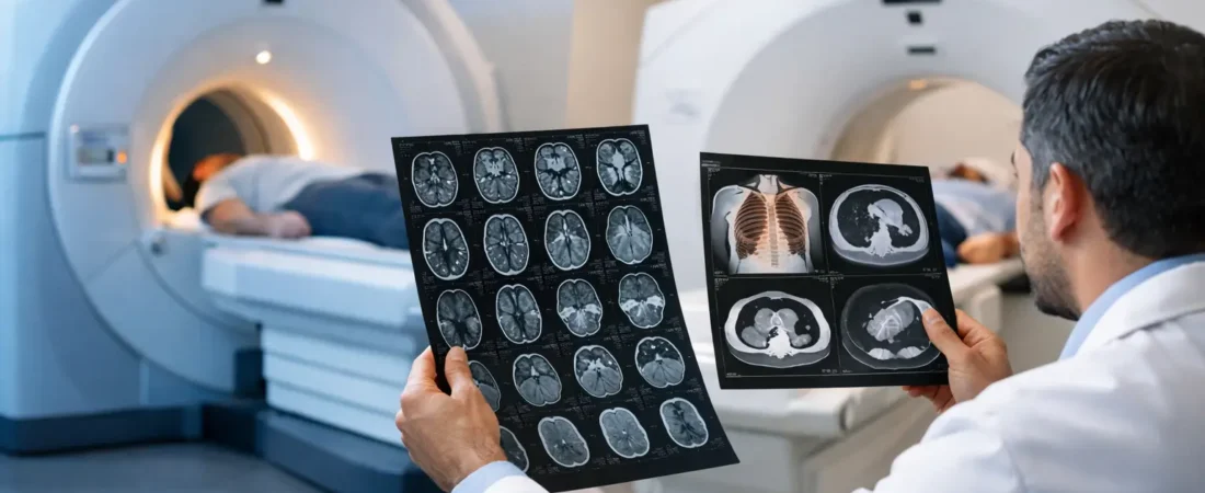
A CT scan, short for computed tomography, uses X-rays taken from multiple angles to create cross-sectional images. It is particularly good at showing bone, air-filled spaces, and fresh bleeding. Modern CT scanners can capture critical images in seconds, a fact that matters when a diagnosis may change treatment immediately.
An MRI, or magnetic resonance imaging scan, uses a powerful magnetic field and radio waves rather than ionizing radiation. Its strength is contrast: it can distinguish among many types of soft tissue with remarkable precision. Brain structures, spinal discs, nerves, ligaments, tendons, muscle, bone marrow, and certain tumors often emerge with far more definition on MRI.
Neither scan is a universal truth machine. Each is an instrument chosen to answer a specific clinical question. The wrong choice may produce a technically excellent image that still leaves the central mystery unsolved.
When CT Is Called First
In emergency medicine, time can be part of the diagnosis. CT often enters the case early because it is fast, widely available, and exceptionally useful for conditions where minutes matter.
Head trauma and suspected brain bleeding
After a serious fall, a car crash, or a blow to the head, clinicians may order a noncontrast CT of the head to look for acute bleeding, skull fractures, swelling, or pressure on the brain. Fresh blood is often conspicuous on CT, making the scan a crucial early tool in suspected stroke or traumatic brain injury.
A CT scan does not rule out every brain problem. A small, early ischemic stroke, certain subtle injuries, and many causes of persistent neurological symptoms can be better seen on MRI. But when hemorrhage must be found or excluded quickly, CT is often the first move.
Fractures, chest injuries, and internal trauma
CT can expose injuries that plain X-rays miss: complex facial fractures, small spinal fractures, pelvic injuries, or damage within the chest and abdomen. In trauma settings, CT may also reveal collapsed lung, internal bleeding, organ injury, or blood around major vessels.
Contrast dye can make blood vessels and organs easier to assess. This is why CT angiography is frequently used when clinicians suspect a pulmonary embolism, aortic emergency, aneurysm, or certain arterial blockages. The image is not merely a picture. It can become a map for urgent surgery, clot treatment, or close monitoring.
Kidney stones and certain abdominal emergencies
The sudden, severe pain of a kidney stone is a familiar diagnostic puzzle. A CT scan is highly effective at locating many stones and identifying obstruction. CT also plays a major role in evaluating appendicitis, bowel obstruction, diverticulitis, perforation, and some cancer staging questions.
The trade-off is radiation exposure. A single CT is often medically justified, especially in an emergency, but it uses more radiation than a standard X-ray. Clinicians weigh that exposure against the risk of missing a dangerous condition. For children, younger patients, and people likely to need repeated imaging, that calculation deserves particular care.
When MRI Finds What CT Cannot
MRI is rarely the fastest option, but speed is not always the priority. When the question involves fine soft-tissue detail, the scan can reveal patterns that CT only hints at – or cannot see at all.
The brain, spinal cord, and nerves
MRI is central to investigating multiple sclerosis, many brain tumors, seizure-related structural abnormalities, pituitary disorders, spinal cord compression, and disc disease. It can detect areas of inflammation, old injury, subtle stroke, and changes in white matter that require interpretation within the patient’s full history.
That last point matters. An MRI can find incidental abnormalities – small changes that are real but unrelated to a person’s symptoms. A frightening phrase in a radiology report is not automatically a diagnosis. Images acquire meaning through the clinical story, examination, laboratory findings, and comparison with prior scans.
Ligaments, tendons, marrow, and joints
A runner with a stress injury may have normal X-rays early on. An athlete with a torn ligament may have a joint that looks structurally intact on CT. MRI is often better at exposing the evidence in these cases: bone marrow edema, cartilage damage, meniscus tears, rotator cuff injuries, tendon pathology, and soft-tissue swelling.
This is also why MRI is commonly used for persistent back or neck symptoms when there are signs of nerve involvement. It can show discs, nerve roots, spinal canal narrowing, and the tissues surrounding the spine in a way CT generally cannot match.
Characterizing masses and inflammation
Both CT and MRI are used in cancer care, but their roles differ by body region and clinical question. CT may be preferred for a fast, broad survey of the chest, abdomen, and pelvis. MRI may better define a liver lesion, pelvic mass, prostate abnormality, breast finding, or the relationship between a tumor and nearby nerves, vessels, or organs.
MRI can also be valuable for infections or inflammatory processes involving bone, joints, the spinal cord, and soft tissues. In suspected osteomyelitis, for example, marrow changes may appear on MRI before they are obvious on other studies.
The Constraints Hidden Outside the Image
The choice is not based on anatomy alone. A CT scan is usually much quicker and more forgiving of movement. MRI takes longer, often 20 to 60 minutes or more, and requires the patient to remain very still. Claustrophobia, severe pain, confusion, or an inability to lie flat can make MRI difficult without additional support.
MRI safety also requires screening. Some implanted devices, metal fragments, aneurysm clips, or other hardware may make MRI unsafe or require special protocols. Many modern implants are MRI-conditional, meaning scanning may be possible under specific conditions, but the imaging team must verify the details. Never assume an implant is safe because it was safe for someone else.
Contrast agents create another layer of decision-making. CT contrast typically contains iodine and can be a concern for people with prior contrast reactions or impaired kidney function. MRI contrast often uses gadolinium-based agents. Serious reactions are uncommon, but kidney function, pregnancy, allergies, and the purpose of the scan all affect whether contrast is used.
Pregnancy changes the equation too. MRI without contrast may be considered when medically necessary, while CT is generally avoided when an alternative can answer the question. Yet a dangerous emergency is still dangerous during pregnancy. When clinicians recommend imaging, they are balancing known risks against the risk of delayed diagnosis.
A Negative Scan Is Not Always the End of the File
One of the most unsettling moments in medicine is being told that a scan is normal while symptoms remain. Sometimes that result is reassuring. Sometimes it means the condition is too early, too small, intermittent, outside the scan’s field of view, or simply not visible with that technique.
A normal head CT may be followed by brain MRI when symptoms point toward a small stroke or inflammatory process. A CT showing a questionable liver finding may lead to MRI for closer characterization. Conversely, an MRI may identify an abnormal area, while CT is used next to evaluate bone involvement, calcification, or the chest and abdomen.
This is not indecision. It is the logic of layered evidence. Imaging works best when the test follows a well-formed question, and when the next question changes based on what the first image reveals.
The Most Useful Scan Is the One That Fits the Clues
CT is often the rapid-response instrument: bleeding, fractures, trauma, lungs, blood vessels, kidney stones, and acute abdominal danger. MRI is often the deeper investigation: brain and spinal cord detail, nerves, ligaments, marrow, soft tissues, and complex lesions requiring sharper contrast.
If severe symptoms are sudden – such as stroke signs, severe chest pain, major trauma, trouble breathing, or a sudden worst-ever headache – seek urgent medical care rather than trying to choose a scan from home. For persistent or unexplained symptoms, the most helpful question for a clinician is not “Can I get an MRI?” It is: “What diagnosis are we trying to find, and which image is most likely to expose it?”
The body does not always leave obvious evidence. But when the right tool is matched to the right clue, the blank space inside a case file can begin to take shape.